

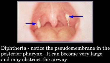
Diphtheria is an upper respiratory tract illness characterized by sore throat, low fever, and an adherent membrane (called a pseudomembrane) on the tonsils, pharynx, and/or nasal cavity. Diphtheria toxin produced by C. diphtheriae, can cause myocarditis, polyneuritis, and other systemic toxic effects. A milder form of diphtheria can be restricted to the skin.
Diphtheria is a disease of the throat, nose, or trachea, caused by a germ called the Klebs-Löffler bacillus. The bacilli produce a white or yellow membrane which is usually plainly visible when it is on the tonsils and surrounding parts, but it may be so thin that it can scarcely be noticeable. If it is in the nose or trachea, its location prevents it from being seen. Other bacteria often grow with the diphtheria bacilli and produce swellings and abscesses. The disease will usually develop within a few hours or days after infection with the germs.
Recognition of Diphtheria
…There are two methods of recognizing diphtheria: 1, by looking into the throat for a membrane; and 2, by taking a culture from the throat or nose.
When a doctor is called to see a sick child, the invariable rule ought to be that he look into the child’s throat. Doctors sometimes yield to the desires of the child or of its parents and do not examine the throat, and thus they often fail to recognize diphtheria in its early stages while it may readily be cured. The presence of spots or a membrane on the tonsils or other situation in the back part of the throat is strongly suggestive of diphtheria, but it is not always a proof of the disease, for they may be due to other causes, such as simple tonsillitis, or septic sore throat, or Vincent’s angina.
Cultures.—The only sure indication of diphtheria is to find diphtheria bacilli in a culture from the throat or nose.
Carriers.—Diphtheria germs may grow in the throat without producing sickness. They usually disappear from the throat in about two weeks after a person recovers from diphtheria. If they persist for three weeks or more, the person is classed as a carrier.
Carriers harbor the bacilli in situations to which the blood-serum is unable to penetrate. The bacilli have been found among the epithelial cells of the tonsils. They may also grow in the crypts of the tonsils and in folds of the mucous membrane of the nose. An abnormal condition of the nose or throat can be seen in nearly every diphtheria carrier, and the germs persist because of the abnormality.
A diphtheria carrier can give the disease to another person. Most cases of diphtheria are caught from unrecognized and unsuspected carriers.
Virulence Test.—Diphtheria bacilli vary in their virulence and in their ability to produce toxin. If a variety has only a slight virulence, it cannot produce the disease in another person, and the carrier is harmless.
A virulence test is performed in the following manner: A culture is taken from a carrier, and the diphtheria germs are isolated from it in a pure culture. A small quantity of the germs are taken and killed and are then injected into the skin of a normal guinea-pig. If the bacilli are virulent, they will produce a red, sore spot in three or four days. The test is like the Schick test on human beings. Laboratories of departments of health are now prepared to make virulence tests on cultures from carriers.
Treatment of Carriers.—A healthy nose or throat will seldom harbor diphtheria bacilli. The procedures which are of value in ridding a carrier of the bacilli are those which would tend to restore the throat to a normal state if no diphtheria germs were present. Most carriers have enlarged tonsils. The removal of the tonsils and adenoids from the throats of those who have them is almost certain to rid a carrier of the germs. Nearly every diphtheria carrier is immune. If he were not immune he would have the disease in an active form. The administration of antitoxin, therefore, has no effect on the bacilli in their throats.
Routine Diphtheria tests were stopped because the majority of people were positive for Diphtheria in the throat and the skin.
Diphtheria infections are further complicated by the toxigenicity factor. If one is infected with a toxigenic strain of C. diphtheriae, two factors must be present to cause the production of the Diphtheria toxin:
-
1. Low extracellular concentrations of inorganic iron. In the presence of iron, a repressor molecule, coded for by the diphtheria toxin repressor (DtxR) gene, is activated by iron and prevents transcription of the tox gene. When concentrations of iron drop, the repressor molecule is inactivated and transcription of the tox gene proceeds, resulting in production of diphtheria toxin.[11]
-
2. Presence in the bacterial chromosome of a lysogenic prophage which contains the tox gene. The bacterium originally receives the tox gene from a specific Beta-phage.[11]
Diphtheria Toxin is a bacterial exotoxin which consists of an active (A) domain, and a binding (B) domain. The toxin acts by binding to the HB-EGF receptor on cells and enters by receptor-mediated endocytosis. The acidic environment of the endosome causes the toxin to undergo a conformational change which causes part of the A domain to enter the endosomal membrane. The A domain is then cleaved from the B domain and passes through the membrane into the cell cytoplasm. The A domain then catalyzes the transfer of ADP-ribose from NAD to Elongation Factor II (EF2), a key protein in protein synthesis during translation. With the addition of ADP-ribose to EF2, EF2 is deactivated. Once a significant amount of EF2 proteins have been deactivated, the cell death occurs.[11]
Diagnosis and Treatment
Clinically, the disease is described as: “An upper-respiratory tract illness characterized by sore throat, low-grade fever, and an adherent membrane of the tonsils, pharynx, and/or nose” [3]
If diphtheria is suspected, then a sample of the bacteria is isolated from the patient and cultured, and toxigenicity testing is performed. If the bacteria is C. diphtheriae, the substrain is further identified – intermedius, mitis, and gravis. To test for toxigenicity, the Elek test is performed. The CDC can also perform a PCR test, but it is not typically used when forming a diagnosis. Another test is serologic testing, which determines the level of antibodies to the diphtheria toxin. [3]
Diphtheria is treated with diphtheria antitoxin, and a 14-day course of antibiotics, preferably Erythromycin or Penicillin.
Is it Diphtheria or something else?
The Acute Infectious Diseases Of Childhood. Diphtheria
This is a disease of the throat. It is caused by the germ that causes diphtheria, that is, by the Bacillus diphtherae. There is no doubt about this. In fact so certain are medical men that this germ causes the trouble that when they fail to find the germ in the excretions (“Bacteriological examination is necessary for diagnosis since some cases cannot be told on inspection alone from acute tonsilitis, and other cases have no membrane at all”—Emerson, Essentials of Medicine), they name the disease something else. The disease may present a perfect clinical picture of diphtheria and no germ be present. This is pseudo-diphtheria and receives another name. One may only have ordinary tonsilitis, “sore throat,” and, if the germ is found, it becomes diphtheria. It was adding thousands of cases of this latter type to the diphtheria figures that enabled them to show a 100% increase in the diphtheria case rate and a corresponding nearly 50% decrease in the death rate, without any lessening of the actual number of deaths, but often with an increase in deaths, when diphtheria antitoxin came into use. The supposed diphtheria germ is often found in the mouth and throat of healthy people who do not have, have not had, and do not subsequently develop diphtheria.
The Encyclopedia Britannica tells us: “If, in diphtheria, the bacillus is not found, the illness is renamed something else.” Sir Wm. Oster, M. D., says in his The Principles arid Practice of Medicine, Page 151, under diphtheria: “The presence of the Klebs-Loeffler baccillus is regarded by bacteriologists as the sole criterion of true diphtheria and as this organism may be associated with all grades of throat affections, from a simple catarrh to a sloughing, gangrenous process, it is evident that in many instances there will be a striking discrepancy between the clinical and the bacterial diagnosis.”
The germ is found in simple catarrhal conditions and also in the mouth and throats of healthy infants and children; and is often absent from the throats of those presenting clinical pictures of diphtheria…
In his Mother’s Hygienic Handbook, 1874, Dr. Trall asserted “the pathological identity of croup and diphtheria.”
“Membranous croup” is the worst form of diphtheria. These cases seldom appear to be very ill. For two or three days there is a rough, croupy cough which becomes a little more croupy each afternoon and evening, but wearing off somewhat in the forepart of the night and in the morning. The child’s breathing is not affected, he has an appetite and there is usually little uneasiness on the part of parents. Then, suddenly, the child almost suffocates. He tosses about on the bed, sits up and struggles in various ways in an effort to breathe. He becomes blue. In severe cases the child suffocates unless relieved by incubation or tracheotomy. In the milder cases the paroxysms are soon over, but they some times recur later…
Formerly croup was divided into membraneous and nonmembraneous or simple croup. Membraneous croup is now regarded as diphtheria. Dr. Trall thought the two croups differed only in degree and said “in the former case the exudation which forms on the mucous lining of the wind pipe (trachea) concretes into a membraneous covering, and in the latter case, the excreted matter is expectorated without consolidation.”
The differences in the behavior of the two exudates show a big difference in their characters, and points to differences in their causes. Simple croup is of a catarrhal nature and results from carbohydrate plethora; membraneous croup is of a serous nature and is the result of protein poisoning. Protein poisoning is more virulent than starch poisoning.
Croup is breathing difficulty accompanied by a “barking” cough. Croup, which is swelling around the vocal cords, is common in infants and children and can have a variety of causes.
…Before the era of immunizations and antibiotics, croup was a dreaded and deadly disease, usually caused by the diphtheria bacteria. Today, most cases of croup are mild. Nevertheless, it can still be dangerous.
Croup tends to appear in children between 3 months and 5 years old, but it can happen at any age. Some children are prone to croup and may get it several times.
In severe cases of croup, there may also be a bacterial super-infection of the upper airway. This condition is called bacterial tracheitis and requires hospitalization and intravenous antibiotics. If the epiglottis becomes infected, the entire windpipe can swell shut, a potentially fatal condition called epiglottitis.
Tonsil and throat infections may be caused by either a virus or bacteria, and can be spread from one person to the other through coughing, sneezing and nasal fluids. In preschool children and infants, the common cold virus or flu virus often causes chronic tonsillitis. In adults and adolescents, it is more likely to be caused by bacteria—the streptococcus, staphylocci, pneumococci, or hemophilus bacteria. In rare cases, the bacteria responsible for scarlet fever, diphtheria and mononucleosis can cause tonsillitis.
Acute infectious laryngitis is a common illness in all age groups and again is caused by viruses, especially influenza A, rhinovirus, and adenovirus. Diphtheria is a rare cause.
The illness is generally quite mild with sore throat, hoarseness, cough, and possibly mild inspiratory stridor. Respiratory distress is rare with the exception of young infants. If diphtheria is the cause, respiratory distress with airway compromise is quite marked secondary to the pseudomembrane blocking the laryngeal inlet . Complete obstruction and respiratory arrest may occur. Fortunately, immunization for diphtheria in the United States has made this disease extremely rare.
At one time, the term croup was primarily associated with diphtheria, a life-threatening respiratory infection. Owing to widespread vaccinations, diphtheria has become rare in the United States, and croup currently refers to a mild viral infection of the larynx. Croup is also known as laryngotracheitis, a medical term that describes the inflammation of the trachea (windpipe) and larynx.
Vincent’s angina is a form of sore throat in which there is usually a membrane resembling that of diphtheria. It usually begins as a small, whitish ulcer upon the tonsils or other part of the throat. The ulcer often extends through the crypts of the tonsils and produces an extensive loss of tissue. The disease is to be suspected when a deep ulcer can be seen in the throat or when the throat remains sore and raw after what was called diphtheria. It is caused by a spirochete which occurs in two forms: (1) a large crescent-shaped organism which stains heavily and unevenly; (2) a long, slender spirillum which stains faintly. A diagnosis may be made by taking a specimen of membrane with a swab, making a smear upon a cover-glass, and examining it at once. Large numbers of both organisms will usually be present in a smear from a positive case. A health officer can make a smear from a suspected case and send it to a laboratory for examination.
Vincent’s angina is not common, but it sometimes occurs in epidemics, and a health officer must keep the disease in mind. A case must be controlled in the same manner as one of diphtheria. Its treatment consists of swabbing the ulcer daily with a 20 to 50 per cent. solution of silver nitrate, and of painting the throat frequently with weaker solutions. A cure is indicated by a healing of the ulcer and by the absence of the organisms from smears.
…a milder tonsil infection due to the presence of organisms called the streptococcus and staphylococcus. But the existence of such an infection shows that the little patient’s throat membranes afford good soil for germs, and so it should be guarded against diphtheria with unusual vigilance. Bad teeth, mouth sores, enlarged tonsils, catarrhal inflammations and other abnormalities in the respiratory tract also predispose to the disease. Susceptibility is increased, again, by measles and scarlet fever. In the majority of cases, the germs first find a lodgment in the tonsils.
Acquired immunity to diphtheria is due primarily to toxin-neutralizing antibody (antitoxin). Passive immunity in utero is acquired transplacentally and can last at most 1 or 2 years after birth…
…the increasing percentage of diphtheria cases in adults suggests that many adults may not be protected against diphtheria…
The vaccine does not prevent carriage. The purpose of the vaccine is to make you immune to the toxins the toxogenic strains release. It is the toxins that can make you sick, and the toxins the bacteria releases that make the tonsils or sinuses, etc, an unfriendly environment to other floral bacteria. This in turn, allows the Diphtheria bacteria to take over and become an acute infection.
Antitoxin does not cure the disease and toxin-antitoxin does not prevent it. Both these foreign proteins are responsible for many deaths in both the well and the sick, and for much other injury short of death.
Although comparatively few who come in contact with this disease develop it, it is considered highly contagious and, due to the contagion-superstition, these cases are quarantined. The writer has never handled but one case and saw this but once. After the quarantine was slapped on the case I handled it over the phone. The child made rapid recovery with no complications or sequelae. otein decomposition and by maintaining good health. Diphtheria is a phase of albumenuria.
The use of Diphtheria toxoid in children began in the 1930’s and 1940’s but uptake remained low. However,
..in the 1930s, a gradual rise in diphtheria incidence to 200 cases per 100,000 in the prewar period occurred in Germany and several other central European countries with partially implemented vaccination programs. The onset of World War II in 1939 and the occupation by German troops of many Western European countries led to the last diphtheria pandemic in western industrialized countries.
The Diphtheria Shift
Diphtheria cases remain isolated, with the last outbreaks reported between 1972-1982. Diphtheria incidence continued to decline steadily throughout the vaccine era in the United States and Western Europe (after the immediate postwar period). Cases of clinical diphtheria became extremely uncommon after the 1970s. Residual indigenous cases have been concentrated among incompletely vaccinated or unvaccinated persons of low socioeconomic status…
A feature of these epidemics concerns the age group; most cases have occurred in adolescents and adults, rather than in children.
Age
When diphtheria was endemic, it primarily affected children younger than 15 years; recently, the epidemiology has shifted to adults who lack natural exposure to toxigenic C diphtheriae in the vaccine era and those who have low rates of receiving booster injections. In the 27 sporadic cases of respiratory tract diphtheria reported in the United States in the 1980s, 70% occurred in persons older than 25 years.
Data from Europe are particularly noteworthy because the childhood immunization rate exceeds 95% in some countries (eg, Sweden), but approximately 20% of persons younger than 20 years and as many as 75% of persons older than 60 years lack the protective antibody….

Infection can occur in immunized, partially immunized, and unimmunized persons. However, disease is usually less severe in those who are partially or fully immunized. Diphtheria is endemic in many parts of the world, including countries of the Caribbean and Latin America. The incidence of respiratory diphtheria is greatest in the fall and winter, but summer epidemics may occur in warm moist climates in which skin infections are prevalent. During the 1990s, large epidemics of diphtheria, primarily in adolescents and adults, occurred throughout Asia, the Middle East, Turkey, Albania, Russia, and the independent countries of the former Soviet Union…
The Russian Experience:
What the Russians found was that with toxic diphtheria it wasn’t the antitoxin antibodies in the blood that was important, but how well the person could initiate the interferon response. If a person had difficulty producing interferon, they would get diphtheria and die regardless of their vaccination status or antibody status. If the person’s nutrition, immunity, and their innate immune system are working correctly, diphtheria isn’t a high risk.
Take a look at these studies. What sense does it make for an immunodeficeint child to have a vaccine for which they many not be able to make antibodies for?
A Russian study- child with an immunodeficiency:
“Thymomegalia is registered in every third child in some regions [of Russia].” In this paper the authors confirm that after DPT-immunization of the children with thymomegalia the anti-diphtheria antibodies is not being produced at all or in an insufficient quantity.” (Source: The insufficiency of the anti-diphtheria antibodies production after immunization with DPT vaccine. Kuz’menko L. G., Arziamova V. V. Nedostatochnost’ produktsii protivodifteriinyh antitel u detei s timomegaliei pri immunizatsii vaktsinoi AKDS. Detskie infektsii (Children infections), 2004, 2(7), с. 24-26.)
Also- Clinical-immunological characteristics of the vaccinal process in children with 1st grade thymomegalia:
“It is known that DPT vaccination even in healthy children not only produces a specific immune response, but causes the allergic reorganization in the body, lowers the specific resistance…
The children with modified reactivity from the high-risk groups react to DPT-vaccination by the long-term suppression of resistance, by developing postvaccinal complications, by defective immune response, by high morbidity…
It was demonstrated the DPT-vaccinations (from the first to the third shot) in the most children with thymomegalia of the 1st grade by their first year of life caused the complicated course of the vaccinal process, namely allergic complications, acute respiratory diseases, the lack or inferior immune reaction to diphtheria or pertussis toxins and enlarging the thymus up to 2nd-3rd grade. The result of the three shots was the factual absence of immunity to whooping cough, low anti-diphtheria and high anti-tetanus… immunity.” (Source: Clinical-immunological characteristics of the vaccinal process in children with 1st grade thymomegalia. Adishcheva N. I. Kliniko-immunologicheskie pokazateli vaktsinal’nogo protsessa AKDS u detei s uvelicheniem timusa I stepeni (Abstract of PhD thesis. Tomsk, 1996, pp. 2 and 24. )
What has some other medical research shown?
Incidence of infectious disease and the licensure of immunobiologics in the
United States. ( Am J Prev Med. 1997 Mar-Apr;13 (2):98-103.Campos-Outcalt D, Aickin M. Department of Family and Community Medicine, Maricopa Health System, Phoenix,
Arizona, USA.)
INTRODUCTION: Our objective was to investigate the relationship of vaccine or
toxoid licensure with the incidence of the target disease in the United States.
METHODS: We used a historical correlational study design with outcome measures of the national incidence and elimination rate of polio, pertussis, diphtheria, and measles as well as the New York City incidence and elimination rate of mumps, rubella, and tetanus.
RESULTS: The licensure of pertussis, measles, polio, mumps, and rubella vaccine was followed by an increase in the elimination rate of disease. The elimination rates of diphtheria and tetanus apparently worsened following the licensure of the respective toxoids.
CONCLUSIONS: Historical data provide evidence of proof of efficacy of mass immunization for measles, polio, rubella, mumps, and pertussis, but not for diphtheria or tetanus.
Diphtheria: changing patterns in the developing world and the industrialized world.
In the past, diphtheria was considered one of the most serious childhood diseases because it took a heavy toll in health and life among preschool-aged children. Prior to the widespread availability of diphtheria toxoid, nearly 70% of cases were in children younger than 15 years of age. In the industrialized countries, immunization against diphtheria became widespread in the 1940s and 1950s. This led to a marked decrease in the incidence of diphtheria. There was also a decrease in circulating toxigenic Corynebacterium diphtheriae organisms, resulting in less natural boosting of antibody levels. This had led to gaps in the immunity of the adult population. Since 1990, diphtheria has made a spectacular comeback in several European countries, with a high proportion of cases in adults…
But recently, several developing countries where coverage has been high for 5-10 years have reported diphtheria outbreaks. These outbreaks have been characterized by high case fatality rates, a large proportion of patients with complications, and their occurrence in both young and older age groups…

The Changing Epidemiology of Diphtheria in the Vaccine Era
(pdf: http://www.journals.uchicago.edu/cgi…e?JID981402PDF )
Patterns of Spread
The diphtheria epidemic in NIS provided important information. First, there was a high proportion of cases among adolescents and adults, especially in Belarus, Russia, Ukraine, and in Baltic States (Estonia, Latvia, and Lithuania), and a lower proportion of cases in these age groups in the southern republics of the Caucasus area and Central Asia. Second, the epidemic began as an urban epidemic, with a progressive transition to include rural areas over time. Third, the epidemic initially amplified in groups with high rates of close contacts (e.g., hospitals, military troops, indergartens, schools), and later, it made a transition to a more generalized epidemic involving socioeconomically disadvantaged groups (e.g., alcoholics).
Changes in Immunity Patterns by Age
Changes in the age-wise distribution of the immunity patterns usually have been explained by the argument that immunization led to a marked decrease in the incidence of the disease and to a subsequent reduction of the reservoir of toxigenic C. diphtheriaeorganisms. In the prevaccine era, exposure to toxigenic strains of diphtheria organisms was common, and this provided natural boosts to the development and maintenance of immunity against diphtheria. Children were susceptible, and most adults remained immune to the disease. However, after immunization of children became widespread, diphtheria became rare, so exposure to these bacteria (and the concomitant natural boost of immunity) become uncommon. If adults do not have natural exposure to diphtheria-causing organisms or receive booster doses of diphtheria toxoid, their immunity induced by childhood immunization wanes, and they become susceptible to the disease [6, 14, 15].
*Blogger Note: Exposure to Diphtheria organisms is still common as Diphtheria bacteria are all around you, all the time. But you won’t find what you don’t test for.
A large body of evidence has documented changes in the immunity levels of various age groups in the pre- and postvaccine eras. In the prevaccine era, when the circulation of C.diphtheriae organisms was common and the prevalence of diphtheria cases was high, natural immunity was acquired by overt or subclinical infection. Most newborn infants passively acquired antibodies from their mothers via the placenta. In 1914 in Vienna [16] and in 1923 in New York City [17], »80% of newborns showed evidence of diphtheria immunity (figure 1). During the first several months of life, this passive immunity waned and was gradually replaced by active immunity, which was acquired through increasing exposure to natural infection.
By 15 years of age, 80% of the children had acquired natural immunity against diphtheria. The rate of acquisition of natural immunity, however, differs from country to country, probably due to differences in the intensity of early contact with diphtheria organisms, overcrowding, sanitation, and hygiene [15].
… In the early 1980s, the lowest levels of diphtheria antibodies in various areas of the Soviet Union were found in persons 20–40 years old [40–42], and at present, this least protected group has shifted to persons 30–40 years old. In other countries, low-level protection was found in persons 40–50 years old in Australia [43], England [44], Germany [45], and Poland [32, 33] and in persons 150 years old in Denmark [46], Finland [29], Sweden [47], and the United States [26].
Changes in the Age Distribution of Diphtheria Cases
When diphtheria was a common disease, it most frequently affected children: At least 40% of diphtheria cases were among children !5 years of age, and some 70% of the cases were among children 15 years of age. This classic pattern of diphtheria cases was seen in many countries, including the United States in 1908–1934 [48], Germany in 1929–1931 [49], and England and Wales in 1936–1937 [50]. Shifts in the age distribution of diphtheria cases has usually been explained by the impact of immunization. However, historical data show that a shift of the disease to older ages began before mass immunization was introduced.
…All these observations suggest that changes in the age distribution of diphtheria cases resulted from factors other than vaccination. Socioeconomic factors, such as a general increase in the standard of living, smaller families, and less overcrowding, created an environment in which children were not subjected to the same intensity of infection in their preschool years as they had been previously. On the other hand, increasing enrollment in schools, summer camps, and meetings of children, adolescents, and adults from different neighborhoods and social backgrounds probably contributed to wider circulation of C.diphtheriae within these age groups. Likewise, migration and displacement of many people during World War II probably enhanced the circulation of diphtheria organisms and contributed to the shift toward more adult cases [15].
Host susceptibility is the key to Diphtheria. Even fully immunized persons can have asymptomatic infections of C. diphtheria and can transmit diphtheria. The vaccine won’t make a bit of difference when it is solely the host factors that will determine the nature of the disease and its outcome. Are you told that those who have adequate iron won’t get Diphtheria because the toxin can’t be produced in the presence of iron? The iron issue has more to do with the link to poverty than any other factor. You can have Diphtheria more than once if the conditions are right.
Clinical diphtheria does not necessarily confer natural immunity.
1990 Case Definition
Clinical case definition
An upper respiratory tract illness characterized by sore throat, low-grade fever, and an adherent membrane of the tonsil(s), pharynx, and/or nose without other apparent cause (as reported by a health professional)
Laboratory criteria for diagnosis
· Isolation of Corynebacterium diphtheriae from a clinical specimen
Case classification
Probable: meets the clinical case definition, is not laboratory confirmed, and is not epidemiologically linked to a laboratory-confirmed case
Confirmed: meets the clinical case definition and is either laboratory confirmed or epidemiologically linked to a laboratory-confirmed case
Comment
Cutaneous diphtheria should not be reported.
1995 Case Definition
The 1995 case definition appearing on this page was re-published in the 1997 MMWR Recommendations and Reports titled Case Definitions for Infectious Conditions Under Public Health Surveillance [MMWR 1997;46(RR10)] (available at http://www.cdc.gov/mmwr/preview/mmwrhtml/00047449.htm). Thus, the 1995 and 1997 versions of the case definition are identical.
Clinical description
An upper respiratory tract illness characterized by sore throat, low-grade fever, and an adherent membrane of the tonsil(s), pharynx, and/or nose
Laboratory criteria for diagnosis
- Isolation of Corynebacterium diphtheriae from a clinical specimen, or
- Histopathologic diagnosis of diphtheria
Case classification
Probable: a clinically compatible case that is not laboratory confirmed and is not epidemiologically linked to a laboratory-confirmed case
Confirmed: a clinically compatible case that is either laboratory confirmed or epidemiologically linked to a laboratory-confirmed case
Comment
Cutaneous diphtheria should not be reported. Respiratory disease caused by nontoxigenic C. diphtheriae should be reported as diphtheria. All diphtheria isolates, regardless of association with disease, should be sent to the Diphtheria Laboratory, National Center for Infectious Diseases, CDC.
What Do They Know and When Did They Know It?
Global Epidemiology of Infectious Diseases
To date, diphtheria has not been a major problem in most developing countries. Immunization for infants and children was introduced with the Expanded Programme on Immunization in the late 1970s. Coverage of infants in developing countries with three doses of DPT vaccine rose gradually from less than 10 per cent in 1974 to 81 per cent in 1995 (Expanded Programme on Immunization 1996). In these countries, the process of maintaining immunity still operates through natural mechanisms, including frequent skin infections caused by C. diphtheriae.
Socioeconomic changes, especially rapid urbanization with migration from rural areas, and sociocultural changes, including improved hygiene and different lifestyles, are changing the epidemiological patterns of diphtheria, so that in some developing countries epidemics are occurring, with more serious faucial and laryngeal forms. Algeria, China, Ecuador, Jordan,
Lesotho, and Yemen have reported diphtheria outbreaks which occurred following a 5 to 10 year period of high immunization coverage. These outbreaks have been characterized by high case fatality rates (CFRs), a large proportion of patients with complications, and occurrence in both young and older age groups (Galazka & Robertson 1995).
Completeness of routine reporting:
The true numbers of diphtheria cases and deaths are unknown. In developed countries, where diphtheria occurs in the form of single imported cases or, as recently in the Russian Federation and Ukraine, in defi nite outbreaks, the reporting may be assumed to be good. In developing countries, however, where the disease is usually endemic, reporting systems are
weak and the impact of immunization on disease incidence is monitored primarily through national incidence fi gures. Such statistics are often inaccurate because they are based on incomplete data gathered by the routine surveillance systems, which are usually hospital-based.
Completeness of reporting depends mainly on two elements. First, the public must have access to health services and use them. Second, the health services must report cases accurately and regularly to the appropriate public health authorities. A study in 13 developing countries that compared survey data with reported data found that only 2 to 5 per cent of tetanus cases and 1 to 26 per cent of poliomyelitis cases were detected and reported through routine surveillance systems (Expanded Programme on Immunization 1982). Overall reporting efficiency for the vaccine preventable diseases is estimated to be less than 10 per cent, although this estimate does not specifi cally address diphtheria case reporting (Expanded Programme on Immunization 1994b). Further efforts are needed to establish highly efficient surveillance systems capable of detecting most cases of targeted diseases.
Immunity to diphtheria and tetanus in inner-city women of childbearing age.
B A Koblin and T R Townsend. Am J Public Health. 1989 September; 79(9): 1297–1298.
Sera were collected between August 1984 and June 1986 from women …the last 20 years have found a higher percent of women susceptible compared to the present study. However, these studies have also reported an increase in the proportion susceptible with age. Millian, et al, found low levels of tetanus antitoxin (<0.01 unitlml) in 25 percent of women aged 20-29 years and 37 percent of women aged 30-39 years. Levels of diphtheria antitoxin <0.02 unit/ml were found in 15 percent of women aged 15-19 years, 19 percent of women 20-29 years, and 24 percent of women 30-39 years…
In the present study, the percent of women susceptible to diphtheria was higher in all age groups compared to tetanus. One reason for this observation may be the use of tetanus toxoid alone as the practice for wound prophylaxis after trauma instead of the combined diphtheria-tetanus toxoids. Another reason is that diphtheria toxoid is less antigenic than tetanus toxoid and therefore the duration of seroprotection following immunization may be less than that observed for tetanus toxoid…
However, the protective level of antitoxin for diphtheria is less well defined than that for tetanus. Outbreaks do occur in communities in which immunization has been extensive and immunization with diphtheria toxoid may modify disease rather than prevent its occurrence. Susceptibility to diphtheria in the older age groups can be a result of lack of exposure to diphtheria in the past, lack of completion of the primary series in childhood, or lack of boosters since childhood…
Diphtheria in the United States, 1971-81.
R T Chen, C V Broome, R A Weinstein, R Weaver, and T F Tsai.Am J Public Health. 1985 December; 75(12): 1393–1397.
General Trends. Diphtheria case incidence remained relatively unchanged from the previous decade until 1975, when the rate began to plummet(Figure 1), reaching a low of two cases (0.01 case per million) in 1980. Mortality paralleled the decline in incidence, including no deaths in 1980. The death-to-case ratio remained close to the historically prevailing 10 per cent until 1976, demonstrating the continued high risk of death associated with diphtheria. It has since varied erratically because of the small number of cases annually….

Carriers
The frequency of diphtheria carriage in the United States is not known accurately. Nontoxigenic isolates and carriers not associated with outbreaks were probably not detected by the surveillance system. The 471 noncutaneous carriers that were reported resembled the cases in age, sex, and racial distribution, but had better immunization status and a lower proportion of toxigenic strains isolated.
Discussion
Diphtheria incidence in the United States has fallen steadily since at least the 1920s (Figure 1). The reasons for the decline before the introduction of diphtheria toxoid immunization are
unclear, but may be related to factors such as diphtheria toxin-antitoxin use and general improvements in socioeconomic factors, nutrition, and housing…
The incidence of noncutaneous diphtheria disease have reached the lowest recorded levels ever in the United States. While the degree of diphtheria underdiagnosis is unknown, the continued decline in incidence is further corroborated by the National Center for Health Statistics mortality data. The decline in diphtheria incidence in the United States during 1971-81 occurred despite serologic studies during the 1970s showing subprotective serum diphtheria antitoxin levels in approximately 25 per cent of the children’0″ and 75 per cent of the adults’2 tested in three US cities. Furthermore, the US Immunization Survey, based on interview question- naires, showed a gradual increase in the percentage of children less than 14 years old not immunized with at least three doses of diphtheria and tetanus toxoids and pertussis vaccine (DTP), from 17 per cent in 1971 to 26 per cent in 1981 ‘for Whites, and from 29 per cent to 47 per cent for non-Whites. Surveys of the actual immunization records of the children in the same age group, available since 1980, showed that 10 per cent of the Whites and 32 per cent of the non-Whites in 1981 were not fully immunized.
The experience with diphtheria immunization indicates that an immunization level of 70 per cent 80 per cent is needed to prevent epidemic spread. However, immunization with diphtheria toxoid is protective only against the phage-mediated toxin, and not against infection by the C. diphtheriae organism. Thus immunized persons have less severe disease when infected, but may remain important as asymptomatic carriers in the transmission of disease. Outbreaks in communities with up to 94 per cent immunization levels have been reported. Therefore, some authors have challenged whether “herd immunity” is applicable to diphtheria.
…Also, as diphtheria incidence decreases, the proportion of the population with natural immunity from prior exposure will also decrease.
Also see: Gross Estimation–Diphtheria Statistics Defy Reality
How Long Does Immunity to Diphtheria Last?
Eli Eichelberger.Am J Public Health Nations Health. 1948 September; 38(9): 1234–1238.
From these figures it becomes obvious that diphtheria can no longer be considered a disease of infancy and childhood alone. Such statistics bear out the conclusions of an editorial in the Journal of the American Medical Association which concluded that diphtheria was increasing and that there was a tendency for the disease to become relatively more prevalent among the older age groups. It appears that our thinking regarding this disease will have to be revised, as well as our evaluation of the duration of diphtheria immunity. If a large per cent of adults are now susceptible, it must be assumed that they must never have been immunized, or if previously immunized,they must have lost their immunity. Consequently, one who thinks of diphtheria as a childhood disease may entirely miss the diagnosis in the adult unless facts regarding this disease are appreciated.
Exposure as a Factor in the Age Distribution of Measles, Diphtheria, and Poliomyelitis
W. Lloyd Aycock.Am J Public Health Nations Health. 1934 May; 24(5): 433–437.

…actual infection in non-immunes produces diphtheria with approximately equal frequency at all ages. However, this does not mean that all infected non-immunes develop the disease, but only that risk of disease in infected non-immunes appears to be equal at all ages….
CONCLUSIONS
The variation in the occurrence of measles, diphtheria, and poliomyelitis in non-immune persons at different ages would appear to be due in part to a difference in the frequency with which persons of different ages are exposed to respective viruses, and in part to a difference in the frequency with which persons of different ages are infected under the same apparent degree of exposure. The differences in the risk of infection at different ages in measles, diphtheria, and poliomyelitis would appear to be due to quantitative or qualitative differences in personal, social or household habits of persons of different ages, which habits constitute sanitary habits in the present connection.

MORTALITY FROM DIPHTHERIA DECREASING
Am J Public Health (N Y). 1926 June; 16(6): 621–622.
THE third annual summary of diphtheria mortality in the large cities of the United States deserves more than a passing mention. It has frequently been stated that while the use of diphtheria antitoxin has greatly lessened the mortality of the disease, the incidence of the disease has not shown any decided decrease. In addition to this every health officer knows that many cases of diphtheria are not treated promptly, and many times even where antitoxin is used, it comes too late to do much good. Every year sees the public becoming more and more educated, and there seems reason to believe that diphtheria antitoxin is being more and better employed. Until 1920 but little improvement could be demonstrated in- the mortality from diphtheria in the majority of cities. From 1910 to 1914 only 13 cities had rates which averaged under 10. In the periods from 1915 to 1919 and 1920 to 1924, only 18 cities had an average rate under 10, while in 1924, 37 cities, and in 1925, 49 cities, had rates under 10. Making the estimate according to the population of the 70 cities studied, 29,243,128 in 1923, and 31,049,595. in 1925, we find that the diphtheria death rate in the former year was 13.12 per 100,OQO against 9.74 for 1925. In spite of an increase of practically 2 million in population, the actual deaths for diphtheria decreased more than 800 for these cities. Perhaps even more encouraging than this figure is the fact that 10 cities, all of considerable size, showed death rates under 4 per 100,000, while 8 cities, including several of the largest in the United States, have shown a continuous decline in diphtheria death rate by 5-year periods from 1-890 to 1924.
F. W. Sears.Am J Public Health (N Y). 1925 February; 15(2): 98–101.
The adult diphtheria previous to the beginning of our work was only a fraction of the total number of cases in the city, while during the past year the adult diphtheria cases have outnumbered both those of the school children and the preschool children. This is similar to what occurred following vaccination against smallpox. Previous to the days of vaccination smallpox was considered to be a children’s disease. Since general vaccination it has become an adult disease. May we not expect that the same thing will occur with general immunization of children against diphtheria ?
DIPHTHERIA MORBIDITY AND MORTALITY
Am J Public Health (N Y). 1924 May; 14(5): 427–429.
Laryngeal diphtheria is still a prime cause of death, and it seems that there are still a few physicians who consider membranous croup one disease and diphtheria another. It is especially difficult to obtain positive swabs from cases of laryngeal diphtheria, but it cannot be too strongly insisted upon that membranous croup is laryngeal diphtheria and should be treated as such. It seems necessary also to insist that in every case of sore throat or nasal discharge in which there is any suspicion or possibility of diphtheria swabs should be taken for laboratory examination.
Reasons for the Failure of Our Methods to Control Diphtheria
J. W. Robinson.Am J Public Health (N Y). 1922 June; 12(6): 497–502.
…quarantine is of no value in the control of diphtheria and only leads to a feeling of security that is false, for it is indeed a sad state of affairs that neither the morbidity nor mortality of diphtheria has declined to any great extent during the last ten years. Figures given by Weaver,” Carey,2 Hull,” and others, prove this…
First, transmission requires a close contact, such as actually coughing in the face while someone is inhaling, drinking out of a cup soon after a carrier has used it, or using some other equally good method to transmit the infection. Second, that the percentage of people of any age group who are susceptible, is much less than ‘tests by the Schick test would indicate. The number of secondary cases in families and’ institutions verifies this statement. There is usually more or less contact in either class of places before a diagnosis is made and precautions are taken. And in private homes, unless the case is removed to a hospital, it is exceptional that isolation of the case is carried out until negative cultures are obtained. And yet, what per cent of the other children contract clinical diphtheria? It is very much less than the percentage of susceptibles as determined by the Schick test would lead us to believe. ‘It is difficult to furnish exact figures, as isolation of some cases has been’ complete, and in others the contacts ‘have each’ received a prophylactic dose of antitoxin…
…occasionally one will carry bacilli for some time and then develop a clinical case, but such cases are uncommon. In this connection one must bear in mind that unless the symptoms are typical of diphtheria they may be due to other causes. A point to which others have drawn attention is the occasional absence of a membrane, and other typical symptoms mentioned in text-books. Occasionally one sees a case of sore throat and fever from which a positive culture of diphtheria bacilli is obtained, and yet no benefit is derived from the use of antitoxin. These cases may well be some of those who are carriers that are always found if cultures are taken, and become sick with tonsilitis or other throat trouble.
…It seems from practical experience, as determined from statistics given by various writers and from analysis of conditions existing at present, and which probably will exist until a far future date, that the morbidity rate of diphtheria will remain practically the same.
…Unless it is carefully explained to those immunized that the protection is of short duration, it may cause them to have a feeling of false security at a future date, and so throw them off their guard as to overlook a diagnosis with a possible fatality as the result. While the danger of anaphylaxis is so small as to be negligible, this does not hold true if it is necessary at a later date to use antitetanic or some other serum when the people may forget to tell the physician or he may not ask them of the use of the diphtheria antitoxin previously. The use of a prophylactic dose does not necessarily prevent them from becoming carriers, and it is always a disappointment to the people and disagreeable to the quarantine officers under those conditions to have to extend the quarantine period after the clinical case has cleared up or been removed to a hospital.
DIPHTHERIA CARRIERS AND THEIR RELATIONSHIP TO MEDICAL INSPECTION OF SCHOOLS
Henry Albert.Am J Public Health (N Y). 1912 October; 2(10): 794–798.
It is very certain that diphtheria has been kept alive for months in institutions and communities by such carriers. Indeed it is quite probable that carriers with either a normal throat or a slightly “sore” throat, are the source of more cases of diphtheria than persons affected by well marked cases of the disease.
…The proportion of children not affected by what is recognized as clinical diphtheria but wbo are diphtheria carriers varies naturally according to the extent of exposure. In 1894, Park* found virulent diphtheria bacilli in about one per cent of healthy tlhroats examined in New York City. In 1907, Fisher,t examining the throats of 4,081 healthy individuals living in the Connecticut Hospital for the Insane, where there were a number of cases of diphtheria, found that 2.08 per cent of them were diphtheria bacilli carriers. It has been shown by the examination of a large number of cases, that when clinical diphtheria is not present in a community about one per cent of the population are carriers of diphtheria bacilli, but that when diphtlheria is present in a conmmunity the percentage is very much higher-ranging from 5 to 10 percent. The percentage of carriers among healthy persons who have been quite directly exposed to infection, is often as high as 50.
William C. Woodward.Public Health Pap Rep. 1901; 27: 238–240.
Diphtheria infection apparently depends on the relation between the virulence of the infecting organisms and the susceptibility of the individual attacked. It involves, therefore, questions of quantity as well as quality of the infection, and of degree as well as character of
personal immunity and vital resistance. Unsanitary conditions tend to diminish the vital resistance of the animal organism and, therefore, make it more prone to succumb to attack. Personal uncleanliness on the part of the patient and of those in attendance on him favors
the spread of the disease by increasing the dissemination of infective particles through the atmosphere. Overcrowding tends to the same end by diminishing the absolute quantity of air in the infected room without a corresponding diminution in the amount of infection, thus increasing the density of the infection in the atmosphere, and increasing the likelihood of those exposed to such atmosphere receiving effectively toxic doses of infection. Unsanitary conditions, including personal uncleanliness and overcrowding, are, therefore, active agents in the spread of diphtheria infection.
The Official Definition of Diphtheria
Hibbert Winslow Hill. Public Health Pap Rep. 1899; 25: 243–248.
In no disease is this lack of definition more evident than in the case of diphtheria. Perhaps no disease has been more discussed, certainly no disease- is better known or understood, yet one hears again and again the question asked, where shall the line be drawn between diphtheria and not diphtheria? This question is asked publicly and privately, by the profession, by health officials and by the laity….
Given a combination of patient and bacillus, a long series of possibilities arise. Were all diphtheria bacilli equally virulent and all patients equally susceptible, such a combination of bacillus and patient would result in a definite set of pathological changes in almost all cases and leave little room for indecision in diagnosis. But because of the wide variations in the degree of virulence of the bacillus and in the degree of resistance of the patient, the pathological changes essential to the disease which are the resultant of these two forces (leaving out of the question for the present the effect of environment or of treatment) may be entirely absent, and when present vary in degree almost infinitely. Nevertheless, certain distinct classes may be recognized, and these classes give the key to the situation. The evidence accumulated in the last few years allows us to believe that we may classify the bacilli into two main groups, virulent and nonvirulent, and patients into two groups also, resistant and non-resistant. The virulence or non-virulence of a given diphtheria bacillus may be determined by animal inoculation. This admits of some discussion perhaps, but for our present purposes, we may accept the test as giving a basis for consideration. The resistance or non-resistance of the patient can be determined only, in any specific case, by the development or non-development of the disease, after infection with a virulent organism. This also is a determinable factor in some cases at least. Granting so much then, four classes of cases arise in which bacilli may be found.
Class 1. Non-virulent bacilli found in the throats of resistant subjects.
Class 2. Non-virulent bacilli found in the throats of non-resistant subjects.
Class 3. Virulent bacilli found in the throats of resistant subjects.
Class 4. Virulent bacilli found in the throats of non-resistant subjects.
Cases falling under classes 1 and 2 are probably rare and are not likely to be found except in epidemics or in other circumstances where wholesale bacteriological examinations are made, because ordinarily there would be nothing to call attention to the subject of such infection.
Class No. 3 in which virulent bacilli are found, producing, however, no lesions, on account of the resistance of the subject, are frequently met with.
Class No. 4 includes all the cases of clinical diphtheria-that is, where pathological conditions are present and are sufficiently marked to call -attention to the patient.
So far then, we have arrived logically at the conclusion that the problem presented is to decide whether “diphtheria” is patient + diphteria bacillus simply, or patient + diphtheria bacillus + typical lesions.
I think it will be admitted that the first combination, patient + diphtheria bacillus alone, does not predicate a disease at all-since the essential element of disease is pathological change-and if that combination is not a disease, it cannot be the disease of diphtheria. Nor
is it logical to assert that patient + diphtheria bacillus + typical lesions necessarly constitutes diphtheria although that formula will cover most cases. The typical lesion which attracts most attention and to which we may confine ourselves for the present purpose is an inflammation of the throat, or more definite still, the presence of a pseudo-membrane. If such inflammation, or pseudo-membrane never occurred except as a result of the presence of the diphtheria bacillus, then patient + diphtheria bacillus + inflammation or membrane would be decisive. To such a combination and to such only could the term “disease of diphtheria” be applied. But both inflammation and pseudo-membrane, indistinguishable in practice at times from those produced by the diphtheria bacillus, are of frequent occurrence-the bacilli nevertheless being absent.
Moreover upon such membranes, often due to streptococci, may be implanted diphtheria bacilli, both non-virulent and virulent forms. Thus the combination patient + diphtheria bacillus + pseudo-membrane may exist, where the pseudomembrane is due to a streptococcus solely. Such a case constitutes a disease certainly, but not necessarily the disease of diphtheria. Lest anyone should object that this is a purely hypothetical case, I may cite the not infrequent finding of diphtheria bacilli in the throats of scarlet fever patients suffering from a streptococcus pseudo-membrane, the diphtheria bacilli producing no appreciable effect whatever. It would be no more logical to say that such a patient was suffering from diphtheria because the combination of patient + diphtheria bacillus + pathological changes (due to the streptococcus) was present than to say that patient + diphtheria bacillus + pathological changes (due to the typhoid organism) constituted diphtheria. It seems plain then that the only possible combination which we can logically recognize as the disease of diphtheria is not patient + diphtheria bacillus + typical lesions, but patient + diphtheria bacillus + lesions, due to the diphtheria bacillus. Given this combination, the additional presence of an active streptococcus in the throat does not invalidate the diagnosis of diphtheria any more than would the additional presence of an active typhoid organism in the intestine.
It is evident that the reasoning here used will apply equally well to any other parasitic disease. My only reason for presenting so obvious a conclusion depends on the confusion which sometimes exists amongst the public, and even the profession, especially when the question of
reporting a doubtful case comes up. Thus, a positive culture is obtained from a healthy throat, or from a throat showing the mildest of lesions, or from a streptococcus throat in scarlet fever or other acute specific in which the previous diagnosis was established and the diphtheria bacillus discovered later shows no clinical indications of activity. Thereupon, the attending physician, and the legal medical officer of the district may be in doubt as to the proper classification of the patient.
To the physician, the recognition of the pathological changes and the determination of their cause is the important matter. To the medical officer, the diagnosis, so far as the patient’s welfare is concerned, is of less importance than the protection of the public health. To the one, the presence, or absence of the diphtheria bacillus is regarded chiefly as an aid to diagnosis; to the other, the presence or absence of the bacillus is the essential point at issue. The physician wishes to fortify his patient against the disease of diphtheria, the medical officer desires to protect the public from the bacillus of diphtheria. If a hypothetical case be considered in which the disease of diphtheria was produced by inoculation with the toxin of diphtheria only, the bacilli of diphtheria being absent, the physician would still have the interest of his patient to consider, while the medical officer might go his way free of responsibility. In the far more frequent cases where the bacillus is present, but the toxin absent or ineffective, it is the physician who is relieved of responsibility, whereas the medical officer’s duty to the public remains to be performed. To apply the foregoing considerations to actual practice, it must be remembered that it is impossible at the present time to observe such rules of procedure as will meet every possible condition here outlined.
For instance, it is usually impracticable, from the executive standpoint, to determine in every case the virulence or non-virulence of the bacilli found by the bacteriologist,-certainly it is impossible to determine it in time to be of service in diagnosis. It is necessary then to assume that all the diphtheria bacilli detected are virulent and experiment confirms the natural expectation that such an assumption is correct in the great majority of cases. Again, in any specific case when the diphtheria bacillus is present accompanied by lesions similar to those usually produced by this bacillus, it is often impossible at present to determine immediately and absolutely that those lesions are due to the diphtheria bacillus rather than to other organisms which may be also present. Here again it is usual to assume that the diphtheria bacillus is the cause of the lesions it accompanies,-and this assumption also is usually true. The evident necessity for making these assumptions in practice, perfectly right and proper as such assumptions are in the majority of cases, is just exactly the necessity which gives rise, in the minority of cases, to misunderstanding and confusion…
Report of the Committee on the Cause and Prevention of Diphtheria
J. J. Kinyoun. Public Health Pap Rep. 1896; 22: 56–67.
The so-called membranous croup is yet, we regret to state, a most serious disturbing factor in our fight against diphtheria. It is with surprise and regret that we see how firmly entrenched in the minds of the medical profession is the idea that croup is a non-infectious disease. It has been demonstrated over and over again, so plainly that he who runs may read that nearly nine tenths of these cases are laryngeal diphtheria. Yet despite all this, death certificates are constantly sent in to the board of health as “croup.” These are the landmarks, or rather, tombstones, marking the foci of epidemics of diphtheria. It is noted with satisfaction, however, that many of our state, provincial, and municipal boards of health are beginning to be emancipated from such ideas, and now regard the terms ” membranous croup ” or ” croup ” and ” diphtheria ” as synonyms, and treat them accordingly.
… It is noted especially that the death-rate from “croup ” in the cities of the United States which have not adopted microscopic examinations, is very high in proportion to the number of deaths from diphtheria, and also in proportion to the population; while on the other hand, the number of cases of death from croup in those places in which the diagnosis is made by the culture test, has fallen, and the number of cases of diphtheria has increased somewhat.
Since the adoption of this method for diagnostic purposes, there has been an increase in the number of cases of diphtheria, due in part to the croup cases, already referred to, being properly diagnosed, and the additional cases of laboratory diphtheria. It has been asserted that on account of including these latter cases the death-rate from diphtheria has not been diminished, and the statements made to the contrary are misleading. It must not be lost sight of that the microscopic examinations have been the cause of eliminating a considerable number of cases formerly classed as diphtheria….
It is to be regretted that the statistics from which these data are compiled are not more complete. Many states have a very imperfect system of their vital statistics, and while it is possihIe to obtain data from the majority concerning the death-rate from diphtheria and croup, it is difficult to obtain those dealing with their morbidity.


As you can see, it has been freely admitted in the medical literature that the vaccine had nothing to do with the decline of Diphtheria or mortality rate. The records show that few children had the vaccine when the decline had already begun! Susceptibility is the key, not whether or not you have had the vaccine, and remains that way to this day.
Filed under: Diphtheria, Trading Places | Tagged: carriers, croup, diptheria, epidemiology diphtheria, epiglottitis, laryngitis, strep throat, tonsilitis, Vincent's angina | Leave a comment »


