Rubella Strains

Rubella


Global Measles and Rubella Laboratory Network, January 2004-June 2005


Seven genotypes and three additional provisional genotypes of rubella virus are recognized by WHO (Figure 2). These genotypes are classified into two clades (i.e., groups of similar genotypes), designated 1 and 2; clade 2 viruses have not been found circulating in the western hemisphere. Although knowledge concerning the geographic distribution of rubella genotypes has progressed substantially since 2003, the genotypes of rubella viruses present in many countries and regions remain unknown.


rubella2


Global strains of rubella virus


Based on analysis of the viral E1 gene, there are two genotypes of rubella virus: Genotype I is present in Europe, North America, and Asia; Genotype II is present in China, Israel and Korea and appears to co-exist with Genotype I viruses in these countries 14. While these genotypes differ by 7-11% at the nucleotide level, they differ by less than 3% at the amino acid level and are of the same serotype. Therefore, the biological significance of the evolution and maintenance of two genotypes is unclear. Following the widespread use of rubella vaccine in developed countries from the 1970s onward, a change in rubella strains occurred in which an

intercontinental genotype clade of Genotype I present in Europe, North America, and Japan replaced the geographic clades from each continent. There are still large areas of the world from which no rubella virus isolates have been analyzed (Africa, Australia, and most of mainland Asia) and thus there could be additional genotypes as well as unrecognized geographic clades of Genotypes I and II. It will be important to obtain a selection of rubella virus isolates from these regions before or concurrent with widespread introduction of rubella vaccination so that the pre-existing endemic virus genotypes can be identified…(Frey TK, Abernathy ES, Bosma TJ et al. Molecular analysis of rubella virus epidemiology across three continents, North America, Europe, and Asia, 1961-1997. Journal of Infectious Disease 1998;178:642-650).


EVOLUTION AND DISTRIBUTION OF RUBELLA VIRUS GENOTYPES


Rubella virus is a sole member of Rubivirus group of Togaviridae and its genotypes has been classified in 2004 at WHO meeting. They are genotype 1B, 1C, 1D, 1E, 1F , 2A and 2B as confirmed and 1a, 1g and 2c as provisional. They have unique chronological and geographical characteristics. Genotype 1a worldwide distributed in 1960s and 70s, however almost disappeared since 1980. Genotype 1B mainly has distributed in Europe and genotype 1C in North and South American continents. Genotype 1D has distributed mainly in Asia and genotype 1F is restricted in China. Genotype 1E looks to be derived from genotype 1D and recently becomes to be predominant circulating one worldwide since 1997. Genotype 1g looks to be derived from genotype 1B and distributes in Europe and Americas. Genotype 2A was restricted in China and 2B in Eurasia and Africa. Genotype 2c was found in Russia. Molecular epidemiological study of rubella virus genotype could reveal the transportation of rubella virus from a country to another country. It will greatly help to make a effective plan of rubella immunization program to eliminate and eradicate for a certain country.

Ancestor dating analysis resulted in 1942-46 for virus strains in genotype 1 and 1840 for those in genotype 2.

By these analysis shift of major prevailing genotype of rubella virus may happened in the history of this diseases, at least from genotype 2 to genotype 1 and genotype 1a to genotype 1E.

As rubella has no relating animal viruses as far as studied, this evolution and emergence is very curious to be known in the future.


Mapping of Genetic Determinants of Rubella Virus Associated with Growth in Joint Tissue

J Virol. 2000 January; 74(2): 796–804. PMCID: PMC111599


Rubella virus (RV), the etiologic agent of German measles, belongs to the family Togaviridae and is the only member of the genus Rubivirus. Natural infection in childhood causes a systemic illness characterized by a short-lived maculopapular rash and mild fever (50). The disease is generally benign, and infection is often asymptomatic. It is the teratogenic potential of rubella that brought the virus to the forefront of public health interests and provided the impetus for isolation of the virus and subsequent vaccine development (35, 49). The current vaccine strain RA27/3 has been very effective in reducing the incidence of congenital rubella syndrome in North America, where it is given to all children between 12 and 18 months of age. However like the wild-type strains and the earlier vaccine strain, HPV77/DE5, it is reported to be associated with acute and late-onset joint and neurological symptoms (20, 46, 47, 50).

The association of RV with acute, transient joint manifestations, after both natural infection and vaccination, has been recognized for many years (14, 23, 34, 46, 47). Rubella-associated arthritis (RAA) is usually short-lived, although a number of patients go on to develop chronic or recurrent pauci- or polyarticular symptoms which can persist for some time (7, 8, 22, 43, 45, 47). Studies to define the mechanism of pathogenesis of RAA have been limited by the fact that humans are the only natural host for RV and there is presently no animal model of infection. However, the frequency and intensity of clinical symptoms reported for wild-type (wt) and vaccine strains correlate directly with the ability of the infecting strain to propagate in organ cultures of human synovial tissue, suggesting that tropism for joint tissue is a measure of viral arthritogenicity (31). Although RV strains are genetically around 98% homologous, they display striking phenotypic variation in growth characteristics and plaque morphology as well as tropism for joint tissue (9, 31). The wt strains, such as Therien, which have the highest association with persistent joint symptoms (30%) (47), commonly replicate to titers of 106 to 107 PFU/ml in organ cultures of human joint tissue, comparable to the yields from the most permissive cell lines for RV. The vaccine strain RA27/3, which is associated with much lower levels of recurrent arthritis (4%) (47), is severely restricted in these cultures and does not attain titers greater than 103 PFU/ml. However RA27/3, like the wt Therien strain, was found to persist in joint culture for over 3 months (31). In contrast, no replication of the European vaccine strain, Cendehill, was detected in human joint tissue in this study. Cendehill strain is reported to have a very low association with acute arthritis and none with chronic joint manifestations (4). These results indicate a correlation between the arthrotropism of a specific RV strain and its ability to induce joint symptoms and lends support to the hypothesis that recurrent RAA is triggered by reactivation of virus which has established a persistent infection in the joint.

The Vaccines:


Soon after the rubella virus was first isolated in tissue culture in 1962, several live-attenuated vaccine strains were developed. HPV-77 (duck embryo), HPV-77 (dog kidney) and Cendehill (rabbit kidney) strains were originally licensed in the USA between 1969 and 1970. These vaccines were replaced in 1979 by RA 27/3 (human diploid fibroblast), which produces a strong immune response (similar to natural infection) of 95% or more. While rubella immunity induced by vaccination has been reported to persist for at least 16 years and probably to be lifelong, other recent data indicate that this immunity may wane after 8 years of age. [3] Rubella vaccine is usually offered in combination with measles (MR) or measles and mumps (MMR) vaccines. [1,5] This combination offers the same high levels of immunogenicity and safety as does its individual components. Most of the currently licensed vaccines are based on a live, attenuated strain of rubella virus known as RA 27/3. The vaccines are administered subcutaneously. To avoid interference with possible remaining maternal antibodies the vaccine is usually given at the age of 12-15 months. Attempts to develop killed virus vaccines or sub-component vaccines against rubella have not been successful…


MERUVAX* II

Wistar RA 27/3 strain of live attenuated rubella virus propagated in WI-38 human diploid lung fibroblasts.

(package insert)


BIAVAX® II (Rubella and Mumps Virus Vaccine Live) is a live virus vaccine for immunization against rubella (German measles) and mumps. BIAVAX II is a sterile lyophilized preparation of the Wistar RA 27/3 strain of live attenuated rubella virus grown in human diploid cell (WI-38) culture

PROQUAD -MMRV(Measles, Mumps, Rubella, Varicella)

Has been suspended.


ProQuad* is a combined attenuated live virus vaccine containing measles, mumps, rubella, and varicella viruses. ProQuad is a sterile lyophilized preparation of (1) the components of M-M-R*II (Measles, Mumps and Rubella Virus Vaccine Live): Measles Virus Vaccine Live, a more attenuated line of measles virus, derived from Enders’ attenuated Edmonston strain and propagated in chick embryo cell culture; Mumps Virus Vaccine Live, the Jeryl Lynn™ (B level) strain of mumps virus propagated in chick embryo

cell culture; Rubella Virus Vaccine Live, the Wistar RA 27/3 strain of live attenuated rubella virus propagated in WI-38 human diploid lung fibroblasts; and (2) Varicella Virus Vaccine Live (Oka/Merck), the Oka/Merck strain of varicella-zoster virus propagated in MRC-5 cells. The cells, virus pools, bovine serum, and human albumin used in manufacturing are all tested to provide assurance that the final product is free of potential adventitious agents.

ProQuad, when reconstituted as directed, is a sterile preparation for subcutaneous administration.

Each 0.5-mL dose contains not less than 3.00 log10 TCID50 (50% tissue culture infectious dose) of measles virus; 4.30 log10 TCID50 of mumps virus; 3.00 log10 TCID50 of rubella virus; and a minimum of 3.99 log10 PFU (plaque-forming units) of Oka/Merck varicella virus.


M-M-R® II (MEASLES, MUMPS, and RUBELLA VIRUS VACCINE LIVE)


Package insert

 Note:  “….contains attenuated live measles and mumps viruses propagated in chick embryo cell culture, plus “the Wistar RA 27/3 strain of live attenuated rubella virus propagated in WI-38 human diploid lung fibroblasts.”(1) Principal studies published in the American Journal of Diseases of Children and the American Journal of Epidemiology, reveal that the rubella strain was cultured from an aborted human fetus.(2,3) In addition, the growth medium for the three live viruses that are needed to produce the MMR vaccine is a buffered salt solution “supplemented with fetal bovine serum.”(4) Other ingredients include sucrose, phosphate, glutamate, recombinant human albumin, sorbitol, hydrolyzed gelatin stabilizer, and approximately 25 mcg of neomycin (an antibiotic).(5) The MMR vaccine does not contain a preservative. In fact, according to the FDA, MMR-II never contained thimerosal, a potentially dangerous chemical used in some vaccines.(6) However, trace amounts of mercury were detected in an earlier MMR formulation.”


Genomic sequence of the RA27/3 vaccine strain of rubella virus. 

Arch Virol. 1997; 142(6):1165-80. PMID: 9229006 [PubMed – indexed for MEDLINE]


The sequence of the genome of the RA27/3 vaccine strain of rubella virus (RUB) was determined. In the process, several discrepancies between the previously reported genomic sequences of two wild RUB strains (Therien and M33) were resolved. The genomes of all three strains contain 9762 nucleotides (nts), exclusive of the 3′ poly A tract. In all three strains, the genome contains (5′ to 3′), a 40 nt 5′ untranslated region (UTR), an open reading frame (ORF) of 6348 nts that encodes nonstructural proteins, a 123 nt UTR between the two genomic ORFs, a 3189 nt ORF that encodes the structural proteins, and a 62 nt 3′ UTR. The 5′ end of the subgenomic RNA was found to correspond to a uridine residue at nt 6436 of the genomic RNA. At the nucleotide level, the sequence of the three strains varied by 1.0 to 2.8%, while at the amino acid level, the sequence varied by 1.1 to 2.4% over both ORFs. The RA27/3 sequence will be of use in identification of the determinants of its attenuation, in vaccine production control and in development of second generation RUB vaccines based on recombinant DNA technology.


A comparative field evaluation of three live, attenuated rubella virus vaccines.

Am J Public Health. 1971 January; 61(1): 152–156.


*Before aborted fetal cell lines were used


rubella-strain


Countries using rubella vaccine, by WHO region, as of December 1999* (pg 69)


Rubella and congenital rubella syndrome: global update


rubella


Name That Strain

Measles


Edmonston strain:

Enders’ attenuated Edmonston strain. Edmonston wild-type (wt) measles virus (Parks et al., 2001). In 1954, the measles virus was isolated from an 11-year old boy from the US, David Edmonston, and adapted and propagated on chick embryo tissue culture (CE). The CE adapted strain, known as Edmonston A, was too virulent for vaccine purposes. The strain was attenuated by means of further passages on CE fibroblasts, resulting in a 2nd generation attenuated virus designated as Edmonston B. Again, the strain was too virulent to be applied on a large scale. Laboratories continued to pass Edmonston B on CE until a 3rd generation of more attenuated strains was developed. These strains, which are known by different names and differ from each other in the number of times the parent strain was passed on CE, provide the seeds for the vaccines now commercially available. The measles vaccines supplied through the World Health Organization’s programme on Immunization in the Americas are prepared from seeds derived from Edmonston B (EPI Newsl., 1980). GenBank Taxonomy No.: 11235


For more information on the various Edmonston strains


Schwarz strain:

Further passages of Edmonston A and B on chicken embryo fibroblasts (CEF) produced the more attenuated Schwarz and Moraten viruses, whose sequences have recently been shown to be identical (Combredet et al., 2003). The Moraten and Schwarz strains are highly genetically related, reflecting their common ancestry and similar passage history, and they are safe and effective for most children. Their use has dramatically reduced the incidence of measles, from over 100 million cases in the prevaccine era to approximately 31 million cases in 1997. However, fatal infections have been documented in immunodeficient children vaccinated with these strains (Valsamakis et al., 1999). GenBank Taxonomy No.: 132487

For more information on the various Schwarz strains


The Measles virus is monotypic, or in other words, there is only one serotype. The genetic differences among the various strains can cause variations in the clinical manifestations of infection. Since the Measles virus is monotypic, immunity against one strain can provide immunity against all strains. Even with various genotypes, the viral genomes have not deviated to produce multiple serotypes.


Vaccines:

The Edmonston strain of measles virus isolated in cell culture in 1954 (7) became the progenitor for many live attenuated measles vaccines such as Moraten, Schwarz, Zagreb and Edmonston B (8). Several measles strains were also used for the development of measles vaccines in other countries such as Russia, China and Japan. All these attenuated strains are used around the world and despite the differences in methods employed for attenuation of the original viruses, which involve various cell culture systems, incubation temperatures, and number of passages, remarkable nucleotide sequence similarity has been found among the strains (9).

A live attenuated vaccine (CAM-70) derived from a Japanese isolate has been used in Brazil since 1982. National vaccination campaigns achieved more than 95% coverage after the Brazilian Measles Eradication Program was established in 1992, although some epidemic outbreaks have occurred after the introduction of the program, mainly in 1997 (2,10)…

CAM-70 vaccine was developed in 1970 from the Tanabe strain in Japan by adaptation to the chorioallantoic membrane (CAM) of chick embryos (12-15). This vaccine has been produced at Bio-Manguinhos/FIOCRUZ since the early eighties by a technology transfer from the Biken Institute, Japan.

Since 1963, when both an inactivated and a live attenuated vaccine (Edmonston B strain) were licensed for use in the United States, both the type of measles vaccine and the recommended age for measles vaccination have changed several times. After 1967 and 1975, the inactivated and the Edmonston B vaccine, respectively, were no longer distributed. A live, further attenuated vaccine (Schwarz strain) was first introduced in 1965, and a similar vaccine (Moraten strain) was licensed in 1968. These further attenuated vaccines cause fewer reactions than the Edmonston B vaccine, yet are equally effective. The Moraten vaccine is the vaccine used currently in the United States.


ROUVAX-Live attenuated vaccine against measles (Schwartz strain)


Each vaccine dose contains:

Lyophilizate:

– Hyperattenuated live measles virus

SCHWARZ strain . . . . . . . . . . . . . . . . . ….. . at least 1000 CCID50*

– Human albumin . . . . . . . . . . . . . . . . . . . . . . . . … q.s. for lyophilization

Solvent:

– Water for injections . . . . . . . . . . . . . . . . . . . . . . . . . . . . . q.s. 0.5 ml

*CCID50 = Cell culture infectious dose 50 percent.

This vaccine contains traces of neomycin.

This vaccine also contains lactose.

This vaccine is in conformity with WHO specifications.


M-M-R® II (MEASLES, MUMPS, and RUBELLA VIRUS VACCINE LIVE)


M-M-R* II (Measles, Mumps, and Rubella Virus Vaccine Live) is a live virus vaccine for vaccination against measles (rubeola), mumps, and rubella (German measles).


M-M-R II is a sterile lyophilized preparation of (1) ATTENUVAX* (Measles Virus Vaccine Live), a more attenuated line of measles virus, derived from Enders’ attenuated Edmonston strain and propagated in chick embryo cell culture; (2) MUMPSVAX* (Mumps Virus Vaccine Live), the Jeryl Lynn** (B level) strain of mumps virus propagated in chick embryo cell culture; and (3) MERUVAX* II (Rubella Virus Vaccine Live), the Wistar RA 27/3 strain of live attenuated rubella virus propagated in WI-38 human diploid lung fibroblasts.


The growth medium for measles and mumps is Medium 199 (a buffered salt solution containing vitamins and amino acids and supplemented with fetal bovine serum) containing SPGA (sucrose, phosphate, glutamate, and recombinant human albumin) as stabilizer and neomycin.


The reconstituted vaccine is for subcutaneous administration. Each 0.5 mL dose contains not less than 1,000 TCID50 (tissue culture infectious doses) of measles virus; 12,500 TCID50 of mumps virus; and 1,000 TCID50 of rubella virus. Each dose of the vaccine is calculated to contain sorbitol (14.5 mg), sodium phosphate, sucrose (1.9 mg), sodium chloride, hydrolyzed gelatin (14.5 mg), recombinant human albumin (≤0.3 mg), fetal bovine serum (<1 ppm), other buffer and media ingredients and approximately 25 mcg of neomycin. The product contains no preservative.


ATTENUVAX® (MEASLES VIRUS VACCINE LIVE)


ATTENUVAX* (Measles Virus Vaccine Live) is a live virus vaccine for vaccination against measles (rubeola).


ATTENUVAX is a sterile lyophilized preparation of a more attenuated line of measles virus derived from Enders’ attenuated Edmonston strain and propagated in chick embryo cell culture.


The growth medium for measles is Medium 199 (a buffered salt solution containing vitamins and amino acids and supplemented with fetal bovine serum) containing SPGA (sucrose, phosphate, glutamate, and human albumin) as stabilizer and neomycin. The cells, virus pools, fetal bovine serum, and human albumin are all screened for the absence of adventitious agents. Human albumin is processed using the Cohn cold ethanol fractionation procedure.


The reconstituted vaccine is for subcutaneous administration. Each 0.5 mL dose contains not less than 1,000 TCID50 (tissue culture infectious doses) of measles virus. Each dose of the vaccine is calculated to contain sorbitol (14.5 mg), sodium phosphate, sucrose (1.9 mg), sodium chloride, hydrolyzed gelatin (14.5 mg), human albumin (0.3 mg), fetal bovine serum (<1 ppm), other buffer and media ingredients and approximately 25 mcg of neomycin. The product contains no preservative.


ProQuad® Measles, Mumps, Rubella and Varicella Virus Vaccine Live

MMRV(Measles, Mumps, Rubella, Varicella) 

Has been suspended.


ProQuad* is a combined attenuated live virus vaccine containing measles, mumps, rubella, and varicella viruses. ProQuad is a sterile lyophilized preparation of (1) the components of M-M-R*II (Measles, Mumps and Rubella Virus Vaccine Live): Measles Virus Vaccine Live, a more attenuated line of measles virus, derived from Enders’ attenuated Edmonston strain and propagated in chick embryo cell culture; Mumps Virus Vaccine Live, the Jeryl Lynn™ (B level) strain of mumps virus propagated in chick embryo

cell culture; Rubella Virus Vaccine Live, the Wistar RA 27/3 strain of live attenuated rubella virus propagated in WI-38 human diploid lung fibroblasts; and (2) Varicella Virus Vaccine Live (Oka/Merck), the Oka/Merck strain of varicella-zoster virus propagated in MRC-5 cells. The cells, virus pools, bovine serum, and human albumin used in manufacturing are all tested to provide assurance that the final product is free of potential adventitious agents.

ProQuad, when reconstituted as directed, is a sterile preparation for subcutaneous administration.

Each 0.5-mL dose contains not less than 3.00 log10 TCID50 (50% tissue culture infectious dose) of measles virus; 4.30 log10 TCID50 of mumps virus; 3.00 log10 TCID50 of rubella virus; and a minimum of 3.99 log10 PFU (plaque-forming units) of Oka/Merck varicella virus.


Killed Measles Vaccine (no longer used) 


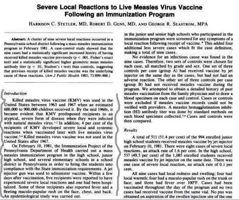
Severe local reactions to live measles virus vaccine following an immunization program.

Am J Public Health. 1983 August; 73(8): 899–900.


killedmeaslesversion


Edmonston-Zagreb (high-titer vaccine)


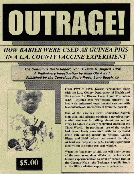
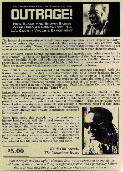
“In an experiment to find out of they could give high-potency Edmonston Zagreb (EZ) measles vaccine to babies as young as four months old [completing disregarding developmental neurology and lack of myelinization in the nervous system of babies] in order to overwhelm their natural maternal antibodies and replace them with vaccine-induced antibodies, medical “researchers” at the CDC and Johns Hopkins University injected thousands of babies in the Third World with the experimental vaccine that reportedly caused chronic immune suppression and the deaths of an unknown number of babies. Also, in the United States, with the help of Kaiser Permanente, more than 1500 six-month old black and Hispanic babies in inner city Los Angeles were “enrolled” in the experiment starting in June 1990. [ During the administration of president and ex-CIA director George Bush.] The study was halted in October 1991, after more than one year of genocidal activity, after repeated reports from vaccine trial sites in Africa that girl babies were dying in higher than expected numbers six months to three years after injection. [A less-than-admirable population control effort.] “–Leading Edge http://www.cco.net/~trufax/vaccine/0696.html   Based on NVIC Vaccine Report 0696 Rec 9/3/96


Also see: MEASLES VACCINE EXPERIMENTS ON MINORITY CHILDREN TURN DEADLY


CDC Admits Informed Consent Violations – CDC director David Satcher admitted in a June 17 Los Angeles Times article that a National Institutes of Health (NIH) investigation of the 1990-91 Los Angeles study found that informed consent regulations had been violated because the parents were not told their babies would be injected with an experimental vaccine that had never been licensed by the FDA for use in America. Both Kaiser and the CDC have denied that any of the Los Angeles babies were harmed by the high potency EZ vaccine but did admit that one child, who received a standard potency EZ vaccine, died from a bacterial infection they maintain is unrelated to the vaccination.


The high potency EZ measles experiments began at four major sites in the mid-1980’s including Haiti, the Senegal, Guinea Bissau and Mexico. Other trials followed in Cameroon, Gambia, Bangladesh, Togo, Iran, New Guinea, Peru, Rwanda, Sudan, South Africa, Egypt, Philippines, Uzbekistan, Thailand, Zaire and Los Angeles. Primary funding came from the U.S. Agency for International Development (USAID) and the World Health Organization (WHO). In Haiti, infants were given the experimental vaccine at 10 to 500 times the usual dose levels. In a June 1996 article in Journal of Infectious Diseases, Johns Hopkins researchers report that infants with the highest antibody responses to high titer measles vaccine have the most profound immune suppression.


Warnings Ignored The measles vaccine experiment was only stopped two years after the director of one of the African sites warned the WHO and CDC experiment leaders in April 1990 that African mortality data raised a red flag about the high titer EZ vaccine. His reports were first ignored and then discounted and he was replaced as a principal investigator. After his mortality data was dismissed as incorrect for more than a year, with support from colleagues at Harvard, he published the mortality data in The Lancet in October 1991. WHO then called for all the sites to submit mortality data for independent analysis. The CDC has stated that enrollment for the LA study was halted in October 1991.


outrage1


outrage2


EDMONSTON-ZAGREB MEASLES VACCINE PROJECT


Studies comparing Edmonston-Zagreb (EZ) measles vaccine with Schwartz measles vaccine in young infants (four to nine months of age) have been conducted in several developing countries, including Guinea-Bissau, Senegal, Haiti, Gambia, Togo, and the Philippines.


These studies revealed high seroconversion rates with EZ vaccine even in the presence of high maternal measles antibody. Several of these studies that used high-dose (4.84 In TCID50) EZ measles vaccine found increased mortality predominantly or exclusively among African female infants immunized with high-dose EZ measles vaccine. In these cases, death occurred approximately one year after vaccination. The causes of these deaths are common causes of infant and child mortality in Third World settings, e.g., respiratory infections, diarrhea and dehydration. Data from these, as well as other studies which did not find higher mortality post-vaccination, were reviewed during a two-day meeting organized by the Expanded Programme on Immunization (EPI), World Health Organization (WHO), in February 1991. Principal investigators of the major clinical trials of EZ vaccine and an international panel of experts attended the meeting and reviewed the data. The panel felt that the probability that the higher mortality was due to the vaccine was very low. Among the reasons cited was the lack of biologic plausibility, particularly in reference to the sex-specific findings.


DIPLOVAX HDC 4.0Freeze-dried measles vaccine produced from the Edmonston-Zagreb strain which has been further attenuated in human diploid cells.


The vaccine contains live, attentuated measles virus of the Edmonston-Zagreb strain, propagated in human diploid cells. Each dose of 0.5 mL contains at least 104 CCID50 (cell culture infective doses) of live measles virus. The vaccine is stabilized with a 10% gelatin-sorbitol stabiliser. The freeze-dried vaccine is reconstituted with 0,5 mL Sterile Water for Injections per dose.


TRESIVAC™


…is prepared from live attenuated strains of Edmonston-Zagreb Measles virus propagated on human diploid cell culture, L-Zagreb Mumps virus propagated on chick embryo fibroblast cells and Wistar RA 27/3 Rubella virus propagated on human diploid cell culture.


The reconstituted vaccine contains, in single dose of 0.5 ml. not less than
1000 CCID50 of Measles virus
5000 CCID50 of Mumps virus
1000 CCID50 of Rubella virus.
Diluent : Sterile water for injection.
The vaccine meets the requirements of USP and WHO when tested by the methods outlined in USP and WHO, TRS 840 (1994).


Measles virus are divided into 21 genotypes


Review of the temporal and geographical distribution of measles virus genotypes in the prevaccine and postvaccine eras   (Virology Journal 2005, 2:87)


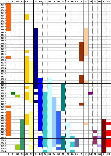
Although measles virus (MV) is serologically monotypic, the genetic characterization of wild-type viruses has identified eight clades (A – H), which have been divided into 22 genotypes and one proposed genotype. Clades B, C, D, G and H each contain multiple genotypes (B1 – 3, C1 – 2, D1 – 10, G1 – 3, H1 – 2) while clades A, E and F each contain a single genotype (A, E, F) [1,2]. The sequences of the vaccine strains indicate that the wild type viruses from which they were derived were all members of genotype A. All measles genotypes can be neutralized by serum from vaccinated persons in vitro, although with varying efficiency [3,4]. There are no known biological differences between viruses of different genotypes. Specific measles genotypes are not associated with differences in severity of disease, likelihood of developing severe sequela such as subacute sclerosing panencephalitis or inclusion body encephalitis, or variability in sensitivity of laboratory diagnosis.

Review of the temporal and geographical distribution of measles virus genotypes 1951 – 2004


Although measles virus (MV) is serologically monotypic, the genetic characterization of wild-type viruses has identified eight clades (A – H), which have been divided into 22 genotypes and one proposed genotype. Clades B, C, D, G and H each contain multiple genotypes (B1 – 3, C1 – 2, D1 – 10, G1 – 3, H1 – 2) while clades A, E and F each contain a single genotype (A, E, F). The sequences of the vaccine strains indicate that the wild type viruses from which they were derived were all members of genotype A.
There are no known biological differences between viruses of different genotypes. Specific measles genotypes are not associated with differences in severity of disease, likelihood of developing severe sequela such as subacute sclerosing panencephalitis or inclusion body encephalitis, or variability in sensitivity of laboratory diagnosis.


Analysis of the variability in the nucleotide sequences of wild-type MVs has enabled the use of molecular epidemiologic techniques for measles surveillance. Genetic characterization of viral isolates or RT-PCR products is the only laboratory test that can differentiate between vaccine-associated cases and wild-type infection.


In 1998, the World Health Organization (WHO) recommended a standard protocol for the designation of measles genotypes. The minimum amount of sequence data required to assign a virus to a genotype are the 450 nucleotides encoding the carboxy terminus of the N protein. The entire sequence of the coding region of the H gene should be obtained from representative isolates.

 
The purpose of this summary is to collate all available reports of MV genotypes and to standardize the published genotype nomenclature, according to the current WHO criteria, with the aim of giving a comprehensive overview of the distribution of MV genotypes in the prevaccine and postvaccine eras.

Summary of distribution of MV genotypes from the prevaccine era to 2004

measlesgenotype

Genetic Diversity of Wild-Type Measles Viruses: Implications for Global Measles Elimination Programs

measlesmap

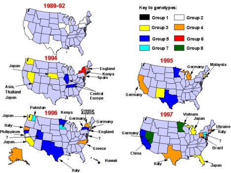
Figure 4. Change in genetic groups of measles viruses associated with U.S. cases and outbreaks between 1988 and September 1997. Arrows indicate sources of virus, if known.


Measles

     Measles was once considered a harmless childhood disease just like Chicken Pox.  You exposed your child to it so they caught it and ‘got it over with’. No one feared measles, just as they did not fear Chicken Pox.

     The Measles vaccine had a low uptake in the past as parents did not want a vaccine for a virus such as Measles because it was considered self-limiting and benign. Children began receiving the vaccine more widely only after the 1977 Childhood Immunization Initiative  and school vaccine mandates were enforced.


     Measles can be a very useful disease in children. They can build a super immune system after having gone through measles. Children with eczema are often cured or relieved of any signs of the condition. Their speech often improves and they go through a maturation process. Many children have been known to make tremendous developmental strides after measles. In the past, when a child was on dialysis, a hospital might have encouraged parents to naturally expose and infect their child with measles because they saw great improvements in the child’s condition.

Even today, the childhood Immunization Initiative is in full force, but it has not stopped Measles from being eliminated.


History

 
     From 1963 – 1967 the U.S. had used the killed Measles vaccine. It had a very low uptake which was a good thing in retrospect as it was a disastrous vaccine. It was made with killed measles virus, which skewed the recipient’s immune systems, making them more susceptible to measles after just two years, but in a new form- “atypical measles”.  It was characterized by pneumonia, high fever, atypical rash and a high fatality rate. It was a disease which could be gotten repeatedly.  The vaccine was quickly and silently removed.


     A new live vaccine was licensed in 1967, but even that was not used extensively.  At first it was to be given to all infants at approximately 12 months of age. Then it was changed to 6 months, especially if there was measles going around. By 1979, they knew they had problems with this one as well. Babies vaccinated at 6 months of age developed what they called an ‘altered immune response’ which resulted in booster shots at 15 months. Nature published an article which showed that babies under one year of age have very different immune functions and responses than adults do, and simply could not handle the measles vaccine given at that age. It caused immune “energy” rather than an “altered” immune response. Again, these issues were kept quiet and uptake continued to be low. Doctors were encouraged not to report measles cases if possible, so that parents wouldn’t lose confidence in the vaccine. Therefore, you would hear terms such as ‘morbilli-like, or “red measles’.


     Since most epidemic outbreaks in the late 1980’s and early 1990’s occurred in  95 – 100% of vaccinated children, a second MMR ‘booster’ vaccine was added to the schedule. By 1990 the actual disease was much rarer, and was simply a continuation of a trend which had been going on right up until the 80’s even in the totally unvaccinated communities. (Clinical Pediatrics). Speaking of Booster shots, do all children need them? No.  The second dose, or booster shot, is to revaccinate the approximated 5% of people for whom the vaccine never worked the first time, also known as primary vaccine failure. That leaves us with roughly 95% getting revaccinated who may never have needed to be. Secondary vaccine failure is due to waning immunity, and even with a second dose schedule in childhood or early adulthood, outbreaks continue to occur in the vaccinated population.


     Health Departments like to say that keeping unvaccinated children away from vaccinated children will protect vaccinated children. They will also say that vaccines protect children. So isn’t that an oxymoron? If vaccines protect, aren’t they already vaccine ‘protected’?  Unfortunately the answer is no. In 1991 over 60% of Measles cases were in vaccinated children, and cases of Measles continue to occur in the vaccinated.


     If anyone should be wary of Measles transmission it is the unvaccinated from the vaccinated. Right in the package insert, it states that MMR vaccinated children can excrete Measles Virus and the Mumps virus into the environment. The Chicken Pox vaccine can also be excreted with the MMR-V or Varicella vaccine. Babies, unvaccinated, the immunodeficient, and even older persons can be at risk from newly vaccinated people. Why aren’t parents being told this?


Detection of measles vaccine in the throat of a vaccinated child.


Mumps vaccine virus genome is present in throat swabs obtained from uncomplicated healthy recipients.


 Some Basic Facts:


     The measles vaccine had nothing to do with the decline in deaths, and has not affected the number of children hospitalized during epidemic years since its introduction.


     Concerning the 1991 USA measles outbreak, over half the deaths were in the vaccinated and most deaths were in immunodeficeint people. (Washington Post. June 14, 1991, BMJ, 11 May, 1991). When news reports talk of Measles reported deaths or more serious injury, why don’t they tell the whole truth?


     In Africa, children who have a natural measles infection have half the asthma, allergies and eczema compared with their vaccinated peers. (Lancet, June 29, 1996) 


     The Germans considered the risks of the vaccine too high given  the fact that deaths and disease severity had decreased without any reference to a vaccine.  
 
     In the pre vaccine era, mothers’ antibodies protected babies for at least a year to a year and a half. Measles was mainly an infection of 5 – 9 year olds and by 15 yrs, 99% had antibodies. Today, adults and infants under one year of age are acquiring Measles which can be very serious.
 
     Vaccinated mothers cannot give protective antibodies to their babies, like Mothers’ who have had naturally acquired Measles, can. Therefore, young babies for whom measles can be more serious are no longer protected.  In the pre-vaccine era, babies rarely got measles before 18 months because maternal antibodies were very high as a result of natural immunity. Today maternal antibodies are generally so low from a vaccine that it simply does not prime the immune system like natural infection will. Babies are at risk of getting measles at younger and younger ages, because maternal antibodies no longer last 15 – 18 months. So if there is even the slightest nutritional or immunological problem, babies will have an increased danger from the measles virus, as there is a difference between the immune system of a baby and a toddler. Vaccinated babies who have maternal antibodies, or people who have measles suppressed with gamma globulin, can have a higher rate of  immunoreactive diseases, sebaceous skin diseases, degenerative cartilage,  bone disease, and certain tumors.  (Lancet, 5 Jan 1985) Also see:
Maternal antibodies interfere with measles vaccination.


     Now think about this…A study published in BMJ years ago found that a select group of children tested, 50% of those with antibodies to measles had never had any clinical disease, and a small subgroup with rising titers also had no clinical symptoms. Non-symptomatic clinical measles was a common entity. This is also shown for Chicken Pox, and several other diseases. To use antibody statistics as proof of either how dangerous or widespread a disease is is a false argument. Measles, like some other diseases, are also dependant upon regular exposure to the bacteria. Which is why in the U.S. Measles is becoming common amongst older adults, who had it clinically as children. Their long term immunity has been jeopardized by the interruption of the bacteria in the environment, so that their levels are no longer automatically boosted every few years.


Measles Basics-


     One sign or symptom specific to measles is Koplik Spots which look like bluish-white grains of salt which can be seen on the inside of the cheek, near the second upper molar, but may also be on the gums anywhere in the mouth.  In the early stages there is also cough, runny nose and fever. This will last for a few days. (Medicine International, 1984, pg 20, Viral Diseases in Man, 83rd Edition, pg 412.)


     The treatment and cure for Measles is called Vitamin A.  As early as 1932, doctors used cod-liver oil to reduce hospital mortality by 57%.  When antibiotics became the timely treatment, Vitamin A was thrown out, up until the mid-80’s that is. Published studies have found that 72% of hospitalized Measles cases in in the U.S. are Vitamin A deficient. The worse Vitamin A deficiency, the worse the complications and the higher the death rate will be. (Pediatric Nursing, Sept/Oct 1996.)


     Measles does not kill children. It is the complications from measles that might attack an already weak immune system. When it knocks down the immune system, the child may become susceptible to other diseases, or develop a secondary infection due to mismanagement of the illness, such as using fever reducing medication, or with a Vitamin A deficiency.  One of the big reasons why third world children suffer from complications of measles and other diseases can be viewed here


     Vaccinations will always be the higher priority. The focus will be on vaccinating as many as they can and fixing the cause of death is secondary to vaccination. If these children were properly nourished and had access to clean water, they wouldn’t be dying. The substitution of vaccination over proper nutrition, sleep, clean water, etc., will not prevent more serious illness or death.


     There will be some who will say the theory of herd immunity is real, that Measles has declined due to a vaccine; deaths have been prevented, etc. However, when you factor in mild and subclinical cases which often are not counted, what have we really prevented? Incidence data ignores these cases which make it appear to be something it may very well not be. What about the number of deaths and injury from the vaccine itself? Maybe a financial cost factor needs to be done between treating naturally acquired Measles vs. the injuries and death associated with the vaccine.


Speaking of which…MMR and MMR-V coming soon!